Using Otoscopes in Ear Examinations
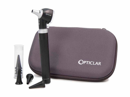
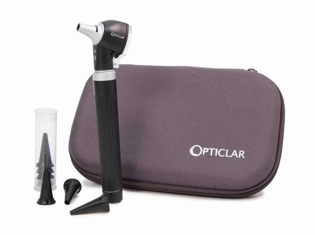
Having the right equipment from a healthcare medical supplier is only part of the task when it comes to offering services to patients. Of course, a comprehensive understanding of how to undertake any medical procedure is essential, such as in the case of using otoscopes to examine the ears. A fibre optic otoscope, or similar device using a type of light bulb, is recommended for most ear canal examinations as it makes the process much easier and more effective when used correctly.
The reason for requiring an ear examination may vary, but typically patients with ear complaints may be unable to provide much specific information about their symptoms, as many different conditions can have similar symptoms while others have none at all. Pain in the ear may also be caused by an entirely different condition, so an otoscopic examination is often used to rule out certain possibilities.
Fortunately, the ear is very accessible and only requires a relatively simple device to examine it thoroughly. This means that these procedures are normally straightforward and can provide information about various factors affecting the ear, including the condition of the tympanic membrane.
The typical process for an examination using an otoscope may be as follows:
1) Examine the skin around the ear and check for any pain or tenderness.
2) Ensure any excess debris, wax or pus in the entrance to the ear canal is removed so as not to hinder the examination.
3) Check the size of speculum which can be fitted to the otoscope and fit into the ear. The largest suitable size will give the best results.
4) Ensure the otoscope is at its default focus setting, which is usually sufficient to complete most ear examinations.
5) Position the otoscope to the ear with your hand, either like a hammer or a pen, making contact with the patient’s cheek to keep the instrument steady even if they move suddenly. It is vital to take care and ensure the speculum does not accidentally cause injury to the ear canal.
Once the otoscope is inserted safely into the ear and correctly aligned, its magnifying lens and light can be used to view the inside of the ear and help to diagnose a range of conditions. It may also be necessary to use the pneumatic function of an otoscope to create small air pressure changes inside the ear and observe the results, in order to diagnose certain tympanic membrane problems.
Recent Posts
-
Cauteries & Cryosurgery for minor procedures
Cauteries and cryosurgery work in a similar fashion, even though they are at opposite ends of the s …4th Jul 2019 -
Choosing the best digital thermometer
As any parent, or healthcare professional knows only too well, getting an accurate temperature read …6th Jun 2019 -
World Hand Hygiene Day
Any healthcare professional will be able to attest to the importance of good hand hygiene. 5 May 2 …1st May 2019

